We are moving our newsletter to Substack for a better experience!
In Week #238 of the Doctor Penguin newsletter, we focus on the recent use of large language models (LLMs) to support and enhance communication in healthcare.
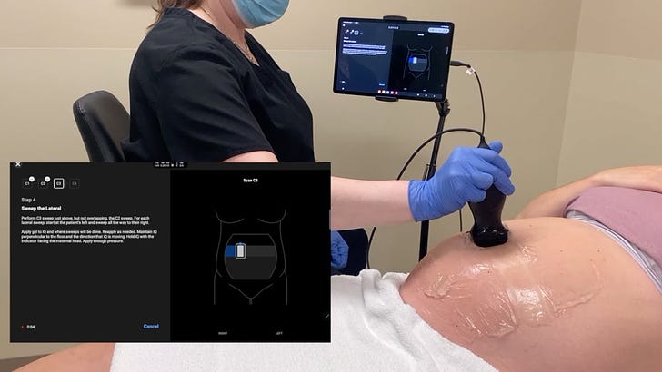
1. Gestational Age. Accurate assessment of gestational age (GA) is essential to good pregnancy care but often requires ultrasonography, which may not be available in low-resource settings. Could AI help novice clinicians accurately estimate GA using a low-cost, battery-powered ultrasonography probe?
Stringer et al. conducted a prospective study evaluating the accuracy of an AI-enabled ultrasound tool for estimating GA when used by novice clinicians with minimal training. The study enrolled 400 pregnant individuals in Zambia and North Carolina and compared GA estimates from novice clinicians using the AI-enabled tool to those from expert sonographers using standard fetal biometry. Between 14-27 weeks gestation, novice clinicians with no prior training in ultrasonography estimated GA as accurately using the AI tool as expert sonographers using traditional ultrasonography devices, with a mean absolute error of 3.2 days vs 3.0 days. Performance remained similar in the 28-36 week window. The AI tool also outperformed last menstrual period dating and fundal height measurements, which are the de facto GA assessment standard in many low- and middle-income countries. However, it was less accurate after 37 weeks. Overall, the results suggest this low-cost, AI-enabled ultrasound device allows minimally trained users to estimate GA as accurately as experts up to 37 weeks, which could expand access to GA dating in low-resource settings.
Read paper | JAMA
2. Ovarian Cancer. Epithelial ovarian cancer (EOC) is one of the deadliest women's cancers. Early detection is key for improving survival (with a 5-year survival rate over 70% in stage I/II compared to 25% in stage III/IV) and can potentially be achieved through methylation markers from circulating cell-free DNA (cfDNA).
Li et al. developed a transformer-based approach to improve early detection of EOC using cell-free DNA. They first pretrained a methylation transformer called MethylBERT on over 110,000 cancer methylation datasets, containing not just EOC but all available cancers, to learn methylome-wide features across different CpG sites (regions in DNA crucial for gene repression and expression). They also surveyed over 3.3 million CpG sites in more than 420 EOC and healthy female pooled cfDNA samples, validating 493 of the most significant methylation markers. Using these markers, they developed a binary classification model called MethylBERT-EOC, which applies the pretrained MethylBERT to methylation data from the 493 selected CpG sites in a patient's cfDNA sample to predict the probability of EOC. The MethylBERT-EOC diagnostic model achieved nearly 90% sensitivity in overall EOC diagnosis, with a significant improvement in early EOC detection, demonstrating 80% sensitivity. This performance was 30-40% higher than traditional biomarkers like CA125 and 13% higher than a conventional LASSO-based model. These results demonstrate significant potential for improving early detection of ovarian cancer through non-invasive blood testing, potentially leading to earlier interventions and better patient outcomes.
Read Paper | Cell Reports Medicine
3. Electrocardiogram. Most clinical diagnoses of cardiovascular disease still rely on the standard 12-lead electrocardiogram (ECG), which requires specialized equipment typically available only in hospitals or clinics. Could AI reconstruct a complete 12-lead ECG from a limited subset of leads, potentially expanding access to this diagnostic tool?
Mason et al. developed a convolutional neural network-based model to reconstruct a full 12-lead ECG using only three leads - two limb leads (I and II) and one precordial lead (V3). Trained on over 600,000 clinically acquired ECGs, the model produced reconstructed ECGs that showed high correlation with the original 12-lead ECGs. A separate classification model for detecting acute myocardial infarction (MI) performed equally well using the reconstructed ECGs compared to the original 12-lead ECGs (AUC = 0.95 for both), providing initial evidence that I + II + V3 leads may be sufficient for identifying acute MI. Notably, when interpreted by cardiologists, the ability to identify ECG features consistent with ST-elevation MI (STEMI) from the synthesized 12-lead ECG (I + II + V3) was not inferior to that obtained from the original 12-lead ECG, with a margin of error of 10%. The study demonstrated that the reconstructed 12-lead ECG is valuable not only for automatic algorithms in identifying acute MI but also for cardiologist interpretation and STEMI identification. Given that the required input can be obtained using commercial sensors without the need for a complete 12-lead ECG setup, this approach may facilitate earlier diagnosis of cardiovascular diseases in remote or resource-limited environments.
Read Paper | npj Digital Medicine
4. Free Flap Monitoring. Free flap surgery is a reconstructive technique that involves transplanting tissue from one part of the body to another to cover a wound or defect, completely detaching the tissue from its original blood supply and reconnecting it to new blood vessels at the recipient site. Frequent postoperative free flap monitoring, such as inspecting flap color and checking temperature, is critical to reduce flap compromise. However, it requires significant clinician time, as it is typically conducted every 2 to 3 hours or even hourly during the early postoperative period.
Kim et al. developed an automated free flap monitoring system to reduce the burden on medical staff. The system comprises two key components: a segmentation model for accurately identifying flaps in images and a grading model for detecting perfusion abnormalities. A smartphone camera with a monitoring application was installed in a place where the flap could be well observed, and the camera automatically took photographs at regular intervals. The system identified the flap area from the images and evaluated its perfusion status. If there were no issues, the process would be repeated. However, if an abnormality was identified, the system would alert pre-assigned medical staff to directly evaluate the flap. The integrated system was successfully applied in a clinical setting for 10 patients, conducting 143 automated monitoring sessions. These results suggest that an automated system may enable efficient flap monitoring with minimal use of clinician time.
Read Paper | JAMA Network Open
-- Emma Chen, Pranav Rajpurkar & Eric Topol

